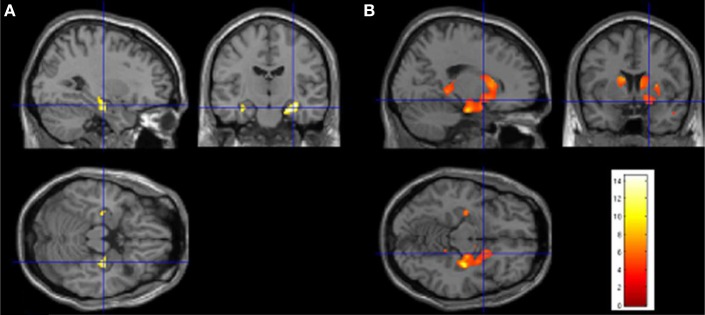
Figure 2.

Quantitative comparison of 3D T1-weighted images of patient ML with 10 age-matched control subjects using voxel-based morphometry (VBM; SPM8, Wellcome Institute, London, UK). For details regarding the method used here, please refer to Labudda et al. (2012). (A) Hypothesis-driven comparison within a hippocampal volume of interest demonstrates a marked reduction of gray matter volume within both hippocampi of the patient (p < 0.05, FWE) which has an anterior and right-sided preponderance (please note that in SPM the right side of the picture is the right side of the patient – see crosshair). (B) Using a whole brain analysis with a less conservative statistical threshold (p < 0.001, uncorrected), there are further reductions of gray matter, affecting amygdalae (bilaterally), bilateral dorsal striatum (mainly caudate and putamen, and to a certain extent the globus pallidus), portions of the ventral striatum (bilaterally), and posterior portions of the pulvinaris complex (also bilaterally), again with a right-sided preponderance.
