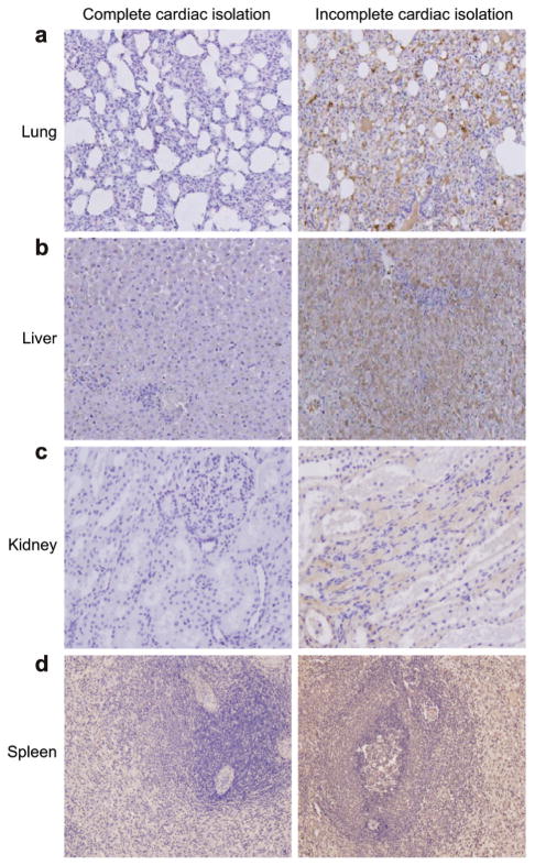
Fig. 3.
Representative green fluorescent protein (GFP) immunohistochemical sections (magnification ×100; ×200) are displayed for MCARD™ with complete and incomplete cardiac isolation. The brown color represents cells with GFP expression and the blue color represents intact cells. Lung (a), liver (b), kidney (c), and spleen (d) cells are shown after histochemical assay for GFP activity. This assay reveals significant collateral organ gene expression after incomplete cardiac isolation. In contrast, there is minimal if any evidence of collateral organ gene expression after complete cardiac isolation. These photomicrographs were taken from animals killed four weeks after the MCARD™ procedure was performed.

