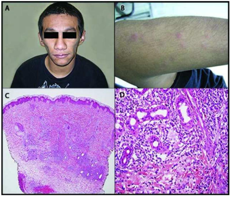
Figure 1.

Proteasome-associated auto-inflammatory syndrome. Clinical photograph demonstrates facial lipoatrophy (A). The patient displayed periodic presence of erythematous papules on extensor surfaces (B). Hematoxylin and eosin stain. Lower magnification demonstrates a perivascular, peri-eccrine, and interstitial mixed infiltrate most prominent in the mid to deep reticular dermis (C). On higher magnification, the infiltrate can be seen to be composed predominately of histiocytes and neutrophils, with admixed lymphocytes and eosinophils (D).
