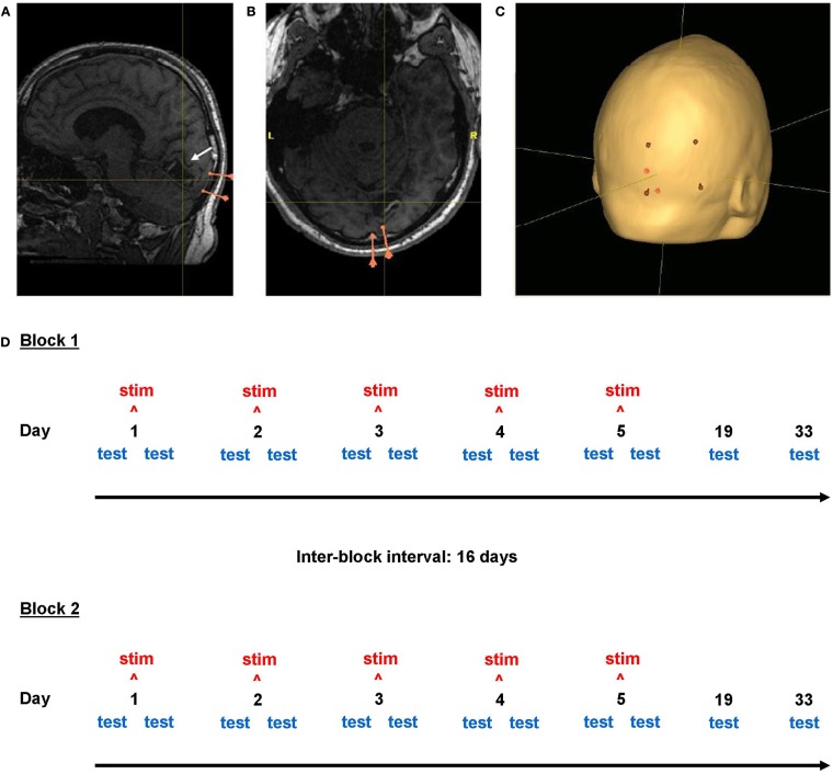
Figure 2.
Neuronavigation and study design. Screenshots from the neuronavigational software Nexstim Eximia. (A,B) Occipital lobe lesion in one subject (center of cross-hairs). Pink markers represent the two meningeal landmarks used for electrode positioning. (C) Scalp localization (red) of the corners of the 5 cm × 7 cm stimulation electrode above the ipsilesional calcarine sulcus (white arrow). Cross-hair location is constant in (A–C). The study design is illustrated in (D).

