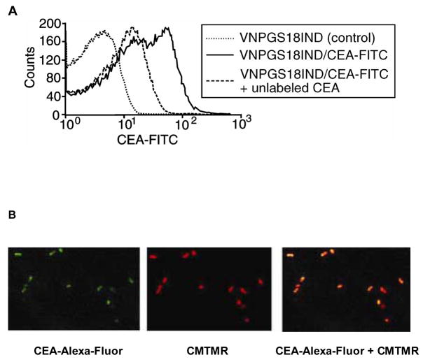
Figure 3. Expression of functional anti-CEA scFv on the surface of VNP/GS18IND.
(A) FACS analysis of CEA-FITC binding to VNP/18GSIND. Bacteria were incubated with medium alone (control, dotted line) or with FITC-conjugated CEA (solid line), or pretreated for 15 minutes with unlabeled CEA and incubated with FITC-conjugated CEA (dashed line). Data are representative of three independent experiments. (B) Confocal images of VNP/GS18IND stained with CEA-Alexa-Fluor (left panel), CMTMR (central panel). The right panel represents the overlay of both stainings. Data are representative of two experiments.

