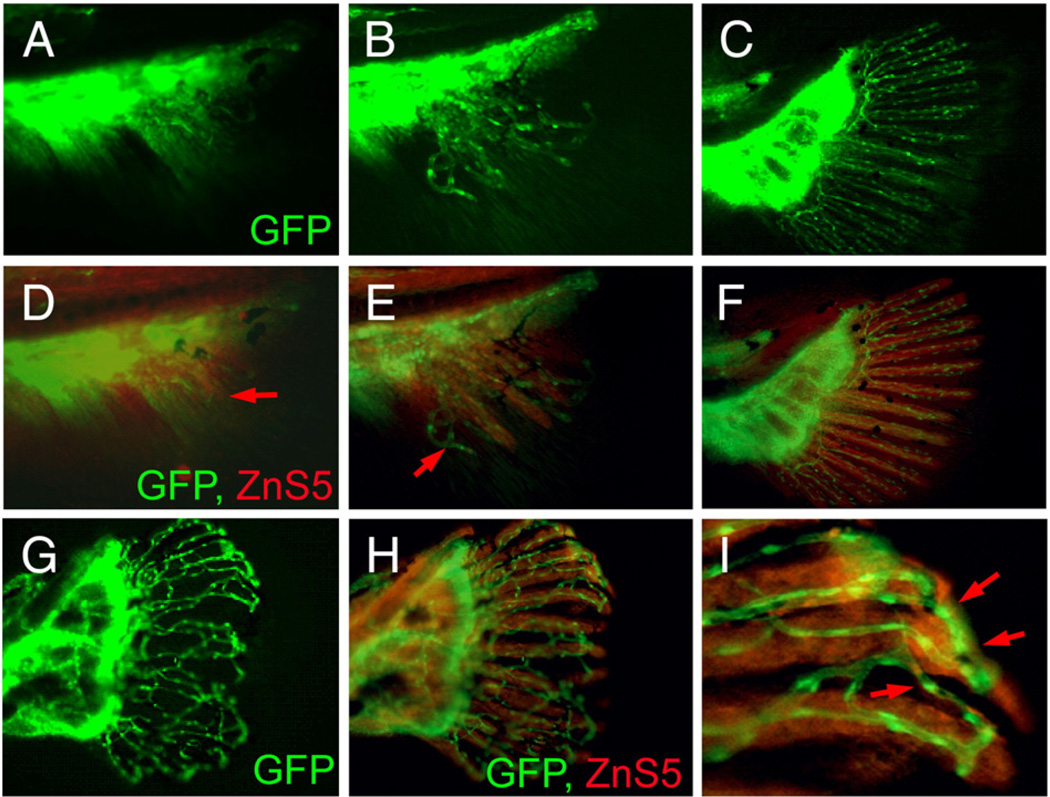
Fig. 7.
Vascular plexus causes wavy and fused fin rays in prp fish. (A–C) Blood vessel development in the caudal fin of wild-type (WT) fish. (D–F) Immunostaining of osteoblasts (red) with the ZnS5 monoclonal antibody of the same fins revealed that osteoblasts developed and were patterned after blood vessels asevidenced byareas where only blood vessels were present (arrows in panels D and E). (G) A prp fin showing the vascular plexus. (H) The same prp fin stained with ZnS5 (red) showing that the osteoblast pattern was closely associated with the blood vessel pattern. (I) High magnification of a representative area of (H) showing wavy and fused osteoblast rays along with the misconnected blood vessels (arrows).

