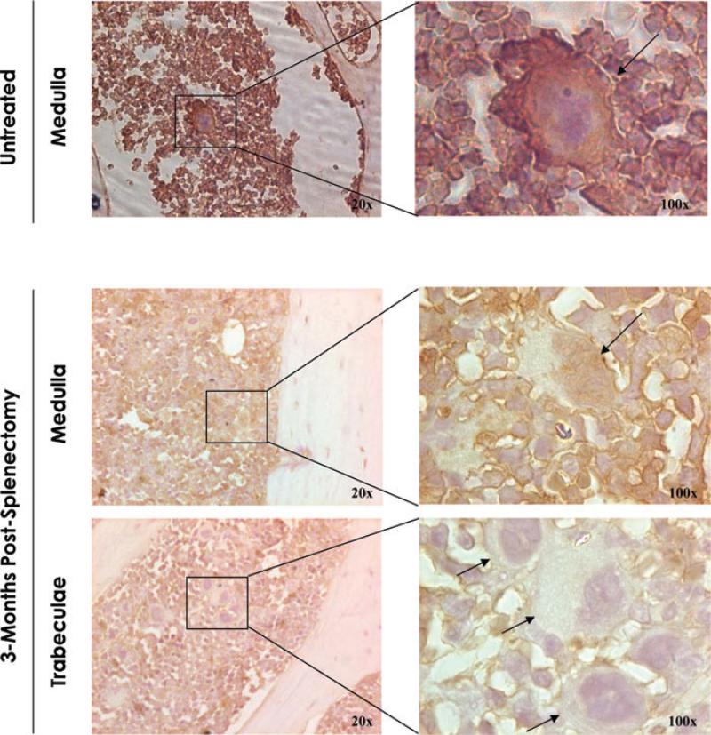
Figure 2.
Megakaryocytes from splenectomized mice express reduced Gata1 content and are localized in areas close to bone trabeculae. Gata1 immunostaining of sections of the long bones from untreated (top panels) and splenectomized (bottom panels) wild-type mice, as indicated. In the case of splenectomized mice, representative areas of the medulla and close to the bone trabeculae are represented independently. In untreated mice, megakaryocytes were detected in the medullary portion of the marrow and were mostly positive for Gata1 (Gata1pos) by immunohistochemistry. By contrast, 3 months after splenectomy, numerous Gata1neg megakaryocytes were detected in marrow sections organized in clusters of cells localized within bone trabeculae. Results are representative of those observed with three additional mice per experimental point. Rectangles indicate areas presented in the enlargements on the right.

