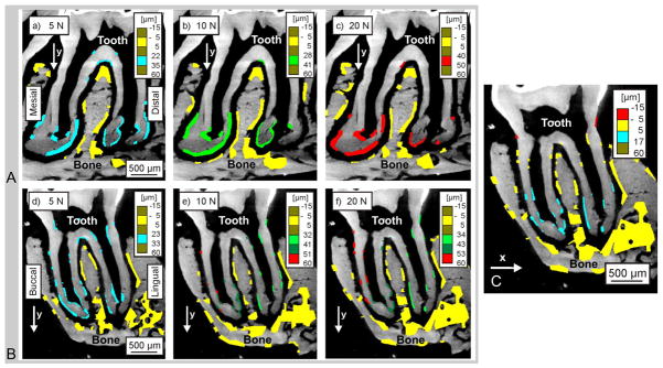
Figure 3. Digital imaging correlation (DIC) used for displacement mapping of the second maxillary molar (tooth) in relation to alveolar bone (AB) at 4x magnification.

2D virtual mesial-distal (A) and buccal-lingual (B,C) sections indicate regions analyzed at 5 N (a, d), 10N (b, e), and 20 N (c, f, and C) of peak reactionary load. All comparisons were made against conditions of no load (0 N). Color-coded regions indicate areas analyzed for changes in horizontal (x shift) and vertical (y shift) displacement fields.
