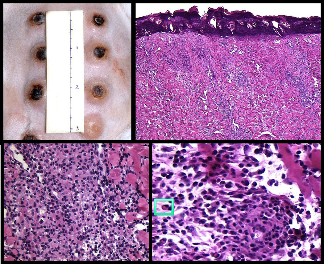
Figure 2. Experimental syphilis infection in the rabbit resembles a human chancre.
Top left panel shows multiple rabbit chancres at the peak of inflammation. Histologically, both the rabbit and human primary chancres have raised, firm erythematous margins with central necrosis (top right panel). Healing occurs within a few weeks in both leaving a small, depressed scar. The inflammatory infiltrate shows a dense, diffuse pattern (bottom left panel) and consists predominately of lymphocytes, macrophages and plasma cells (insert, bottom right panel).

