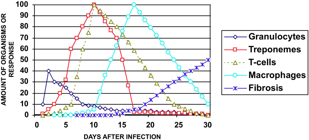
Figure 4. The natural history of a syphilitic rabbit chancre.
This chart illustrates the sequential events in the evolution of the lesions of experimental syphilis. Polymorphonuclear cells are seen in the first few days after inoculation of organisms and then decline. However, in some studies polymorphonuclear cells are seen in higher numbers later during development of the lesion. T. pallidum may been found in very small numbers within 1 day after injection, then increase rapidly so that by day 10–11 the dermis is filled with organisms. T-cells appear within the first few days and increase to a peak about 10 days after infection. Intact organisms co-exist with T-cells during the first 11 days. After 10 days macrophages increase rapidly and by day 13 fragments of digested organisms may be found within macrophages. Organisms are then rapidly cleared from the tissue, so that by 21 days few, if any, may be found. The lesion then heals with regional fibrosis.

