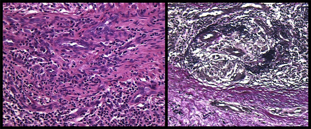
Figure 7. Syphilitic endarteritis obliterans.
Proliferation of swollen, vacuolated endothelial cells accompanied by vessel wall thickening is a frequent finding in cutaneous lesions of syphilis. The inflammatory host response directed at inter-endothelial (see figures 6 and 11) and intra-endothelial spirochetes (90)), likely induces intimal proliferation with subsequent luminal narrowing and ischemia (left panel). Elastic tissue stain demonstrates disruption of an internal elastic lamina and replacement of the intima by fibrous tissue and small vessels (right panel).

