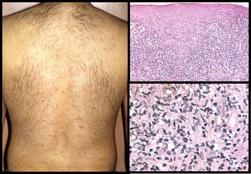
Figure 9. Pustular Secondary Syphilis.
So called rupial syphilis refers to crusted papular and pustular lesions (top left panel). This patient’s histology shows a pustular psoriasis-like pattern with spongiform pustule formation and psoriasiform epidermal hyperplasia (top right and bottom left panels). The presence of plasma cells in the dermal infiltrate is a clue to syphilis (bottom right panel).

