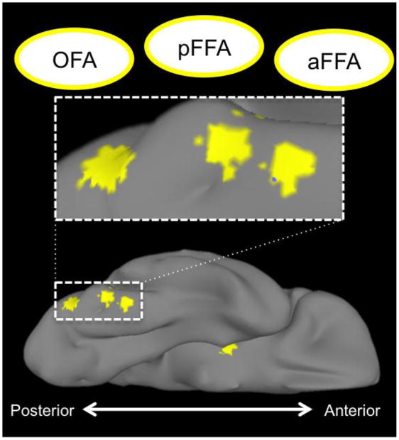
Figure 2. Trio of face-selective regions on the right ventral surface.
Spheres, centered on local maxima locations derived from the group-level GLM, rendered on the surface of the right ventral temporal cortex. These areas include the ‘rOFA’ (most occipital of the three), a posterior fusiform face area, and an anterior fusiform face area. Also visible is the local maxima in the right amygdala.

