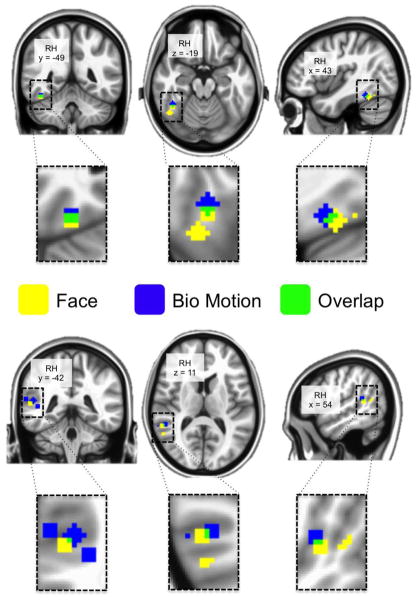
Figure 7. Overlap of peak face- and bio- voxels.
Painted on the cortical surface are spheres centered on local maxima locations of derived from the group-level GLM (see Figure 1) on the right ventral temporal cortex. YELLOW indicates voxels included in a face-voxel sphere, BLUE indicates voxels included in a bio-voxel sphere, and GREEN indicates voxels that are included in both a face- and bio-voxel sphere. The peaks were identified from within our ventral and lateral temporal cortices masks. On the ventral surface there is one bio-voxel peak, which overlaps with the more anterior of two face-voxel peaks. On the lateral surface there are 4 bio-voxel peaks and 2 face-voxel peaks, one of which partially overlaps with one of the bio-voxels.

