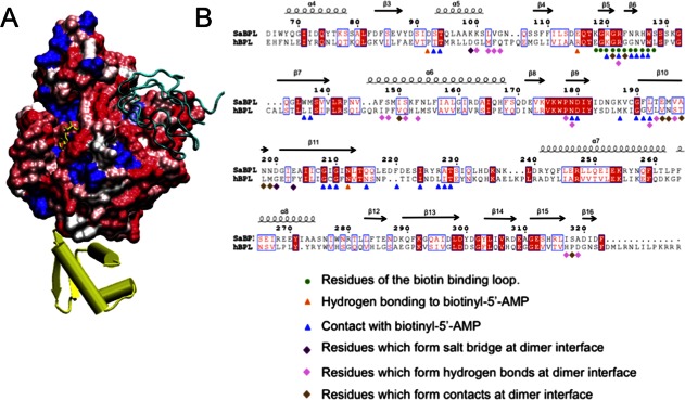
Figure 4.

SaBPL as a potential drug target. (A) Similarity of surface amino acid residues of human BPL mapped to the surface of SaBPL. Identical amino acids are shown in blue. Sequence similarity is shown on a ramped scale from blue through white to red scored using the BLOSUM60 matrix where white indicates sequence similarity, pink and dark pink represent lesser similarity, and red represents the highest level of dissimilarity. The N-terminal DNA-binding domain is completely unrelated to the N-terminal domain of human BPL and is shown in yellow. SaBCCP modeled in its bound position is shown in cyan ribbons. (B) SaBPL sequence alignment with human SaBPL used for the surface comparison. Indicated are SaBPL amino acid residues involved at the catalytic site or the dimer interface of SaBPL.
