
Figure 6.
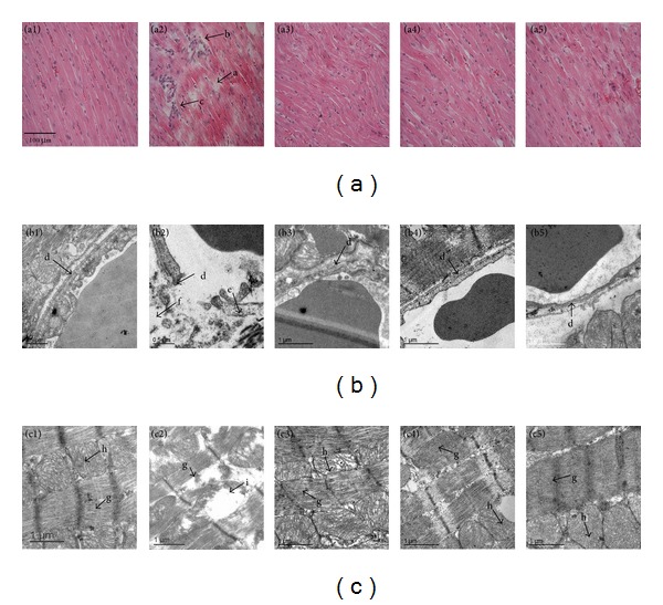
CP pretreatment diminishes I/R-induced alteration in myocardial tissue morphology. (a) Representative images of myocardial sections stained with HE. a: Disrupted myocardial fiber. b: Interstitial edema. c: Inflammatory cell infiltration. Bar = 100 μm. (b) Representative electron micrographs of myocardial capillary from various groups. d: Vascular endothelium. e: Caveolae. f: Interstitial edema. (c) Representative electron micrographs of myocardial fiber in different groups. g: Myofilament. h: Mitochondria. i: Corrupted mitochondria. Results are presented for Sham group (1), I/R group (2), CP 0.1 + I/R group (3), CP 0.4 + I/R group (4), and CP 0.8 + I/R group (5).