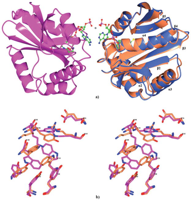
Figure 4.
(a) Ribbon diagram of the Ta0374 dimeric complex with acetyl-CoA/CoA is shown in light blue (molecule A) and magenta (molecule B). The acetyl-CoA/CoA is shown as a ball-and-stick model (nitrogen in blue, oxygen in red, carbon in green, sulfur in yellow). Ni2+ is shown as a grey dot between 3′-phosphate of acetyl-CoA/CoA molecules. Superposition of (A) molecule of the Ta0374 dimeric complex with acetyl-CoA/CoA (light blue) with molecule of the Ta0374 in complex with acetyl-CoA (coral); (b) Stereo view of the superposition of side chain residues that have different positions between (B) molecule (magenta) of Ta0374 dimeric complex with acetyl-CoA/CoA and the molecule (coral) of Ta0374 in complex with acetyl-CoA.

