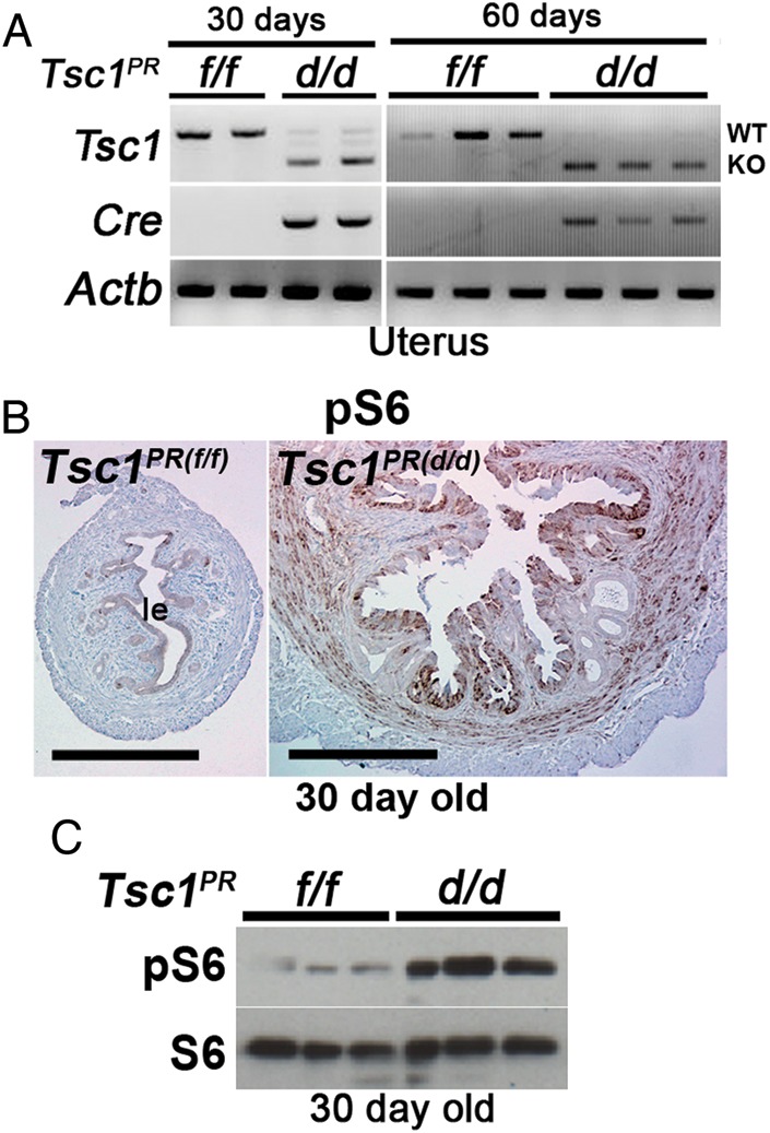
Figure 3.

Heightened mTORC1 signaling in Tsc1-deleted uteri. (A) RT–PCR showing inactivation of Tsc1 in Tsc1PR(d/d) uteri compared with Tsc1PR(f/f) uteri from mice at 30 or 60 days of age. Actb served as a loading control. (B) Immunohistochemistry of pS6 shows increased mTORC1 signaling in Tsc1PR(d/d) uteri (n = 4 females) compared with control Tsc1PR(f/f) uteri (n = 4 females) from mice at 30 days of age. Bars, 400 µm. le, luminal epithelium. (C) Western blotting of pS6 indicates increased mTORC1 signaling in Tsc1PR(d/d) uteri from mice at 30 days of age. S6 served as a loading control.
