Figure 4.
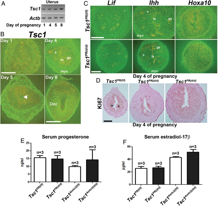
Uterine receptivity is compromised in Tsc1PR(d/d) uteri. (A and B) RT–PCR and in situ hybridization of Tsc1 in wild-type peri-implantation uteri. Tsc1 expression is primarily seen in the epithelium and myometrium (myo) on Day 1, glandular epithelium (ge) on Day 4 and in the stroma (s) on Days 5 and 8 of pregnancy (n = 1 per day of pregnancy). Magnification of Days 1, 4, and 5 of pregnancy: bar, 500 µm; Magnification of Day 8 of pregnancy: bar, 1 mm. Arrowhead denotes the site of blastocyst. Actb served as a loading control. le, luminal epithelium; Dec, decidua. (C) In situ hybridization of Lif, Ihh and Hoxa10 expression Tsc1PR(f/f) (n = 3 females) and Tsc1PR(d/d) (n = 2 females) uteri on Day 4 of pregnancy show comparable expression between the two groups. Upper bar, 250 µm; lower bar, 500 µm. (D) Normal Ki67 staining is seen in Tsc1PR(f/f) females Day 4 (n = 3, left panel), while uteri of two of three Tsc1PR(d/d) females showed compromised epithelial differentiation and stromal proliferation (middle panel); uteri from one Tsc1PR(d/d)mouse showed a relatively normal expression pattern (right panel). Le, luminal epithelium; s, stroma. Bar, 400 µm. (E and F) Serum levels of E2 and P4 on Day 4 of pregnancy (mean ± SEM). Three independent samples were examined in each group.

