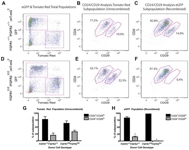
Figure 6.
Quantitative analysis of stem cell populations in FGFR1+/+/R2+/+; mT-mGfl and FGFR1fl/fl/R2fl/fl; mT-mGfl mammary outgrowths using flow cytometry. (A): Magnetic bead, lineage-reduced wild-type FGFR1+/+/R2+/+; mT-mGfl outgrowths were separated into mT-mG-recombined eGFP+ and mT-mG-unrecombined tomato red+ populations to be analyzed separately. (B, C): In wild-type FGFR1+/+/R2+/+; mT-mGfl mammary gland outgrowths, the mT-mG recombined, eGFP+ population and unrecombined mT-mG tomato red+ population demonstrated similar levels of MRU-containing, Lin−CD24+CD29hi cells. Percentages of each subpopulation represent the average between all runs. (D): Magnetic bead, lineage-reduced floxed FGFR1fl/fl/R2fl/fl; mT-mGfl outgrowths were separated into mT-mG-recombined eGFP+ and mT-mG-unrecombined tomato red+ populations to be analyzed separately. (E, F): In floxed FGFR1fl/fl/R2fl/fl; mT-mGfl mammary gland outgrowths, the mT-mG recombined, eGFP+ population contained dramatically reduced levels of Lin−CD24+CD29hi, MRU-containing cells as compared to the mT-mG unrecombined tomato red+ population. Percentages of each subpopulation represent the average between all runs. (G): mT-mG unrecombined, tomato red+ cells from both the wild-type FGFR1+/+/R2+/+ and floxed FGFR1fl/fl/R2fl/fl; mT-mGfl mammary glands did not show significantly different levels of MRU-containing Lin−CD24+CD29hi population. (H): mT-mG recombined, eGFP+ cells from the floxed FGFR1fl/fl/R2fl/fl; mT-mGfl mammary glands show a significant reduction in MRU-containing Lin−CD24+CD29hi cells as compared to mT-mG recombined eGFP+ wild-type FGFR1+/+/R2+/+; mT-mGfl (*, p < .04, n = 2). Graphs represent mean ± SEM. Abbreviations: FGFR, fibroblast growth factor receptor; GFP, enhanced green fluorescent protein.

