Figure 5.
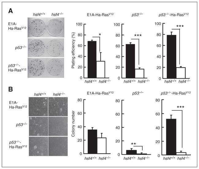
Loss of hsf4 in MEF reduces anchorage-independent cell growth. A, colony formation assay: 102 MEF cells from the indicated genotypes were plated and cultured for 10 days. Colonies were stained by crystal violet, and the total number of colonies containing 50 cells or more were quantitated. Left panel shows representative culture plate for the indicated cell lines. Right panel shows average plating efficiency (number of colonies counted/number of cells plated) calculated from 3 independent experiments. Data represent mean ± SD. P values were determined using 2-tailed Student t test.*, P < 0.05, ***, P <0.001. B, soft agar assay: 2 × 10⁴ cells of the indicated genotypes were plated and cultured on a plate containing 0.7% base agar and 0.35% top agar at 37°C for 14 days. Total number of colonies was quantified. All experiments were repeated at least 3 times. Left panel shows representative plates with colonies formed on soft agar. Right panel shows average colony numbers quantified per plate averaged from 3 independent experiments. Data represents mean ± SD. P values were determined using 2-tailed Student t test; **, P < 0.01; ***, P < 0.001.

