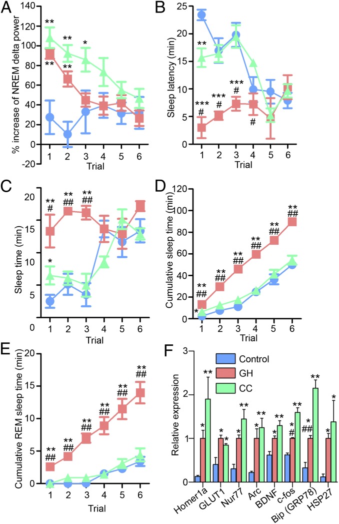
Fig. 1.
Dissociation of delta power and sleep latency following SD by two different methods. NREM delta power (A), sleep latency (B), and total and REM sleep times (C–E) during MSLT following 6-h SD. Mice were deprived of sleep from ZT0–6 by either GH or CC. The control group was allowed to sleep freely during ZT0–6. MSLT, performed from ZT6–9, was composed of six repeats of 30-min trials (i.e., a 5-min period of forced wakefulness followed by a 25-min spontaneous sleep period). EEG/EMG monitoring was performed continuously from ZT0–9. (F) Previously described SD-inducible transcripts were increased in correlation association with delta power as an index of homeostatic sleep need after 6-h SD at ZT6. RNA was extracted from whole brain at ZT6. Values were normalized to Cyclophilin B mRNA levels and expressed relative to the means of the GH group. In Figs. 1–3, data represent means ± SEM, *P < 0.05, **P < 0.01 compared with the control group; #P < 0.05, ##P < 0.01 between the GH and CC groups by ANOVA followed by Tukey’s test (n = 3–7). Arc, activity-regulated cytoskeletal-associated protein; BDNF, brain-derived neurotrophic factor; GLUT1, glucose transporter 1; HSP27, heat shock protein 27.

