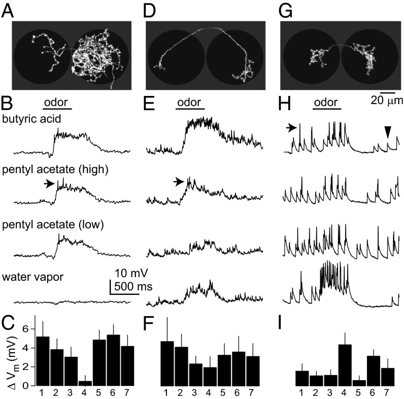
Fig. 2.
Morphology and physiology of Glu-LNs. (A) Morphology of a Glu-LN, shown as a z-projection of a traced biocytin fill. This neuron innervated many olfactory glomeruli, and this pattern was seen in 11 of 29 filled cells. Note innervation of both antennal lobes (black circles), which is typical of Glu-LNs. (B) A whole-cell current clamp recording from a Glu-LN of this morphological type. The spikes fired by this cell (arrow) are small. (C) Mean responses of all of the Glu-LNs of this type (±SEM across experiments), quantified as the change in membrane potential averaged over the stimulus period. Odors are 1, butyric acid; 2, pentyl acetate (10−2); 3, pentyl acetate (10−6); 4, water; 5, methyl benzoate; 6, 1-butanol; 7, ethyl acetate. (D) This neuron innervated mainly a single ventral olfactory glomerulus on both sides of the brain. A similar pattern was seen in 8 of 29 fills. (E) A recording from this type of neuron. Spikes (arrow) are small. (F) Mean responses for all of the Glu-LNs of this type. (G) This neuron innervated the putative hygrosensitive/thermosensitive glomeruli just posterior to the antennal lobe. A similar pattern was seen in 10 of 29 fills. (H) A recording from this type of neuron. Note prominent spikes (arrow) and large excitatory postsynaptic potentials (arrowhead). (I) Mean responses for all of the Glu-LNs of this type.

