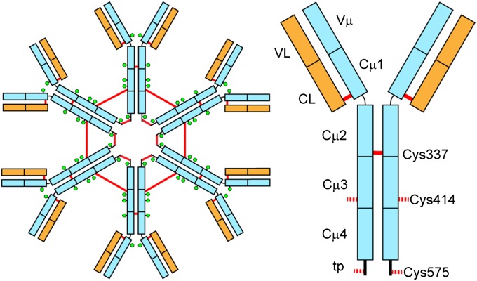
Fig. 1.
Schematic view of hexameric IgM (Left) and a single subunit (Right). Heavy chains are depicted in light blue and light chains in orange. Red lines represent intersubunit disulfide bridges and the green hexagons show glycosylation sites. The cysteine residues that form interdomain disulfide bridges between the Cµ2 domains (C337) and covalently link the IgM hexamer in Cµ3 (Cys414) and the C-terminal tp (C575) are depicted.

