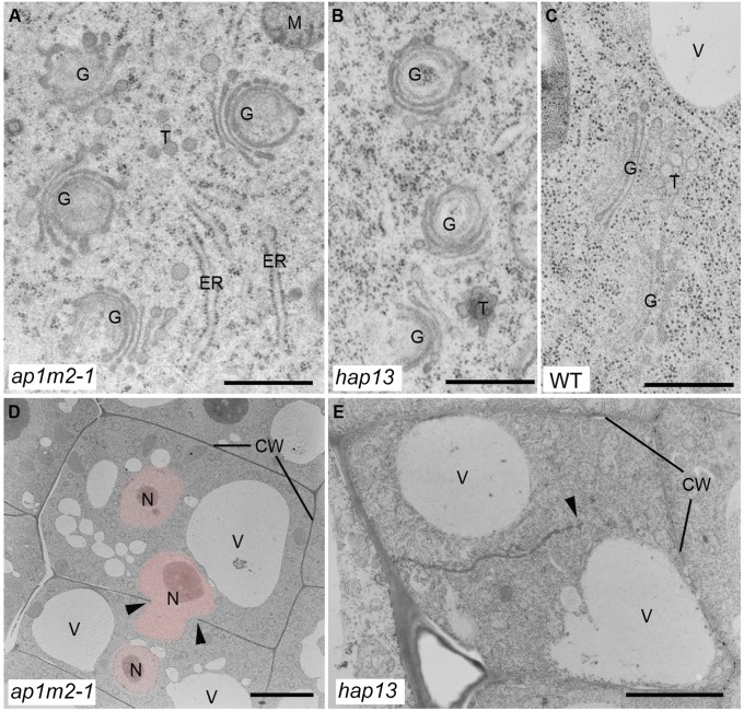
Fig. 5.
Ultrastructural analysis of ap1m2 mutants. Ultrathin sections of cryofixed and freeze-substituted root tips of 5-d-old ap1m2-1 and hap13 seedlings were examined by transmission electron microscopy. ap1m2 mutants displayed bent Golgi stacks (A and B) (compare with WT (C)] and cell wall stubs/incomplete cell walls (arrow heads) (D and E). Note also the presence of several nuclei (pink-colored) per cell in the ap1m2-1 mutant (D). CW, cell wall; G, Golgi stack; M, mitochondrion; N, nucleus; T, trans-Golgi network; V, vacuole. [Scale bars: 500 nm (A–C); 6 µm (D); 3 µm (E).]

