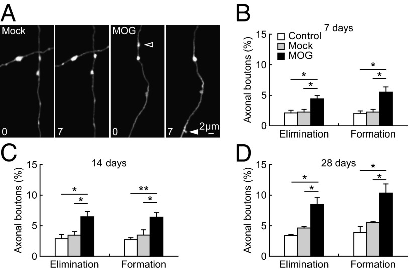
Fig. 3.
Increased turnover of axonal boutons after MOG immunization. (A) Repeated imaging of axonal segments in the somatosensory cortex over 7 d in Mock- and MOG-immunized animals (4-mo-old). Filled and empty arrowheads indicate axonal boutons that were formed and eliminated between the two views. (B–D) Percentage of axonal boutons eliminated and formed over 7 d (B), 14 d (C), and 28 d (D) in no-injection control, Mock-immunized, and MOG-immunized animals. The turnover of axonal boutons was significantly increased in MOG-immunized animals compared with no-injection and Mock-immunized control in all of the time intervals. Data are presented as mean ± SEM. *P < 0.05; **P < 0.01.

