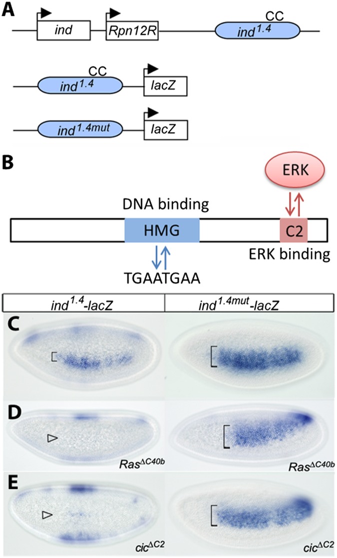
Fig. 2.
Regulation of ind expression by Cic. (A) Diagram of the ind locus. The coding region and its 1.4-kb enhancer (in blue) are separated by the unrelated regulatory particle non-ATPase 12-related (Rpn12R) gene. “CC” indicates a pair of conserved Cic DNA binding sites (TGAATGAA). Reporter genes ind1.4-lacZ and ind1.4mut-lacZ containing WT and mutated CC sites are represented. (B) Schematic representation of the Cic protein highlighting its functional domains. The high mobility group box region binds to the octameric TGAATGAA sites in target gene enhancers, resulting in their transcriptional repression. The C2 motif functions as a docking site for ERK and mediates down-regulation of Cic in response to RTK-dependent ERK activation. Expression of ind-lacZ reporters. In situ hybridization of ind1.4-lacZ and ind1.4mut-lacZ in WT (C), RasΔC40b (D), and cicΔC2 (E) embryos. All images correspond to lateral surface views taken at mid- to late nuclear cycle 14. (C) In the WT background, ind1.4mut-lacZ expression is expanded compared with ind1.4-lacZ expression. (D and E) ind1.4-lacZ appears strongly repressed in RasΔC40b and cicΔC2 embryos (open arrowheads), whereas ind1.4mut-lacZ is insensitive to Cic and EGFR signaling.

