Figure 2.
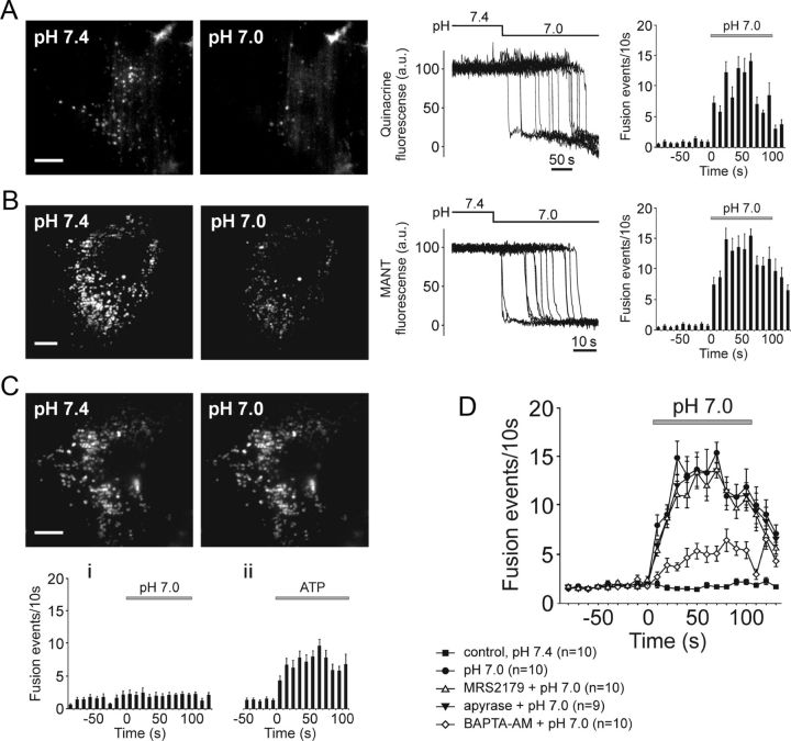
Ventral brainstem astrocytes respond to acidification with an increased rate of exocytosis of putative ATP-containing vesicular compartments. A, B, TIRF images (left) and plots of TIRF intensity changes (middle) showing loss of quinacrine (A) or MANT-ATP (B) fluorescence from a proportion of labeled organelles in cultured ventral brainstem astrocytes in response to a decrease in external pH from 7.4 to 7.0. Plots on the right depict averaged temporal distribution of acidification-evoked fusion events detected in 10 quinacrine loaded (A) and 10 MANT-ATP loaded (B) pH-sensitive brainstem astrocytes. Scale bars, 10 μm; C, Higher basal (at pH 7.4) rate of vesicular fusion events in cultured cortical astrocytes is not affected by acidification. Plots on the bottom depict averaged temporal distribution of fusion events detected in cortical astrocytes exposed to (i) acidification of the external milieu (n = 10 cells) or (ii) application of ATP (10 μm; n = 8 cells). Scale bar, 10 μm. D, Summary data illustrating averaged temporal distributions of acidification-evoked fusion events detected in MANT-ATP-loaded brainstem astrocytes in the absence and presence of MRS2179 (30 μm), ATP degrading enzyme apyrase (25 U ml−1), or after 1 h incubation with a Ca2+-chelator BAPTA-AM (30 μm).

