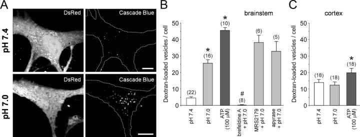
Figure 3.
Ventral brainstem astrocytes respond to acidification with an enhanced endocytotic recovery of the granules. A, Confocal images of cultured DsRed-expressing brainstem astrocytes in control conditions (pH 7.4) and after stimulation with pH 7.0 (10 min) in the presence of 3 kDa dextran conjugated to Cascade Blue. B, Summary data showing the number of fluorescent puncta in the cytosol of ventral brainstem astrocytes stimulated with ATP or exposed to a decreased pH (7.0) in the absence and presence of brefeldin A (50 μm), MRS2179 (30 μm), or ATP-degrading enzyme apyrase (25 U ml−1). *p < 0.01 compared to the number of fluorescent puncta in control conditions at pH 7.4; #p < 0.01 compared to the number of fluorescent puncta at pH 7.4 and pH 7.0. C, Summary data showing the number of fluorescent puncta in the cytosol of cortical astrocytes exposed to a decreased pH (7.0) or stimulated with ATP. *p < 0.01 compared to the number of fluorescent puncta in control conditions at pH 7.4.

