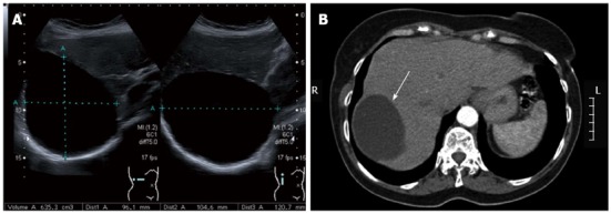
Figure 2.

Simple cyst. A: On abdominal ultrasonography. Ultrasonography (USG) demonstrating a large simple cyst occupying the right hepatic lobe. Note the sharp and smooth border, oval shape, and anechoic echo pattern with the absence of septations and strong posterior wall echoes. The cyst size is indicated by the dotted lines; B: On abdominal computed tomography. Computed tomography demonstrating a sharply defined homogeneous hypodense cystic lesion (arrow) occupying the right hepatic lobe, which was diagnosed as a simple cyst.
