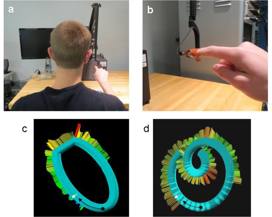
Fig. 1.
Experimental setup. The finger sled attachment (red) is viewed from the back of the subject (panel a) and from the subject’s left side (panel b). Subjects had eyes closed during each trial and their computer monitor was always switched off. Fingertip start position within virtual tubes (black dots), movement direction (black arrows), and contact force direction (white arrows) are indicated for example shapes from experiment 1 (panel c) and experiment 2 (panel d). These screenshots (seen only by the experimenter) also indicate the magnitude of the perpendicular contact forces (by color and length).

