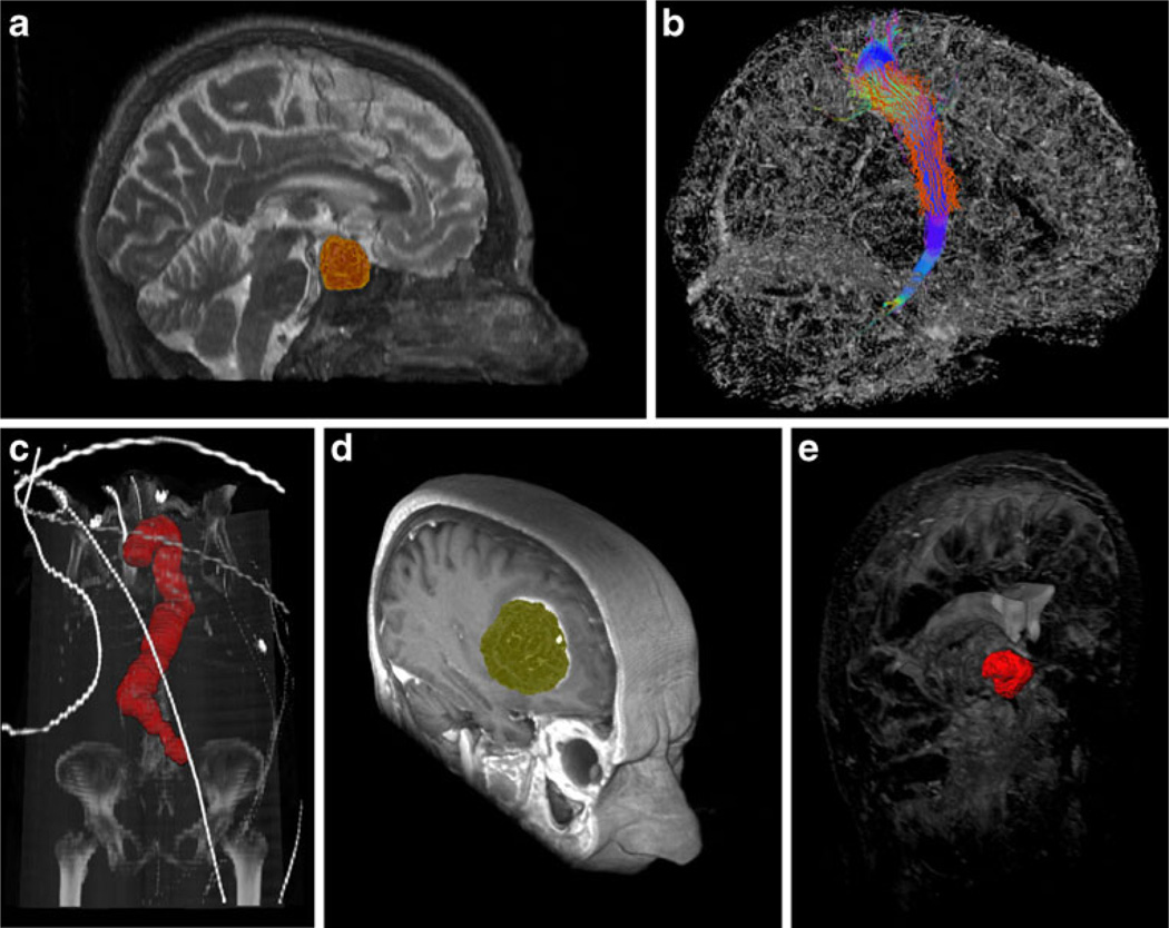
Fig. 1.
Examples from medical applications where we have used graph-based image segmentation approaches. Upper row from the left: pituitary adenoma (MRI) (a), fiber tracts (DTI) (b). Lower row from the left: aorta with abdominal aneurysm (CTA) (c), glioblastoma multiforme (MRI) (d), cerebral aneurysm (CT) (e)

