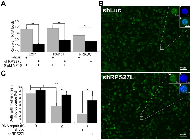
Figure 4. Effects of RPS27L in DNA repair of VP16-induced double-strand breaks in LoVo cells.
(A) Relative expressions of DNA repair genes. The mRNA levels of E2F1, RAD51, and PRKDC were quantified using qRT-PCR following cells with indicated VP16 treatment. The relative expression of each gene was calculated by comparing that of shLuc cells without VP16 treatment. (B) Representative photos of cells with γ-H2AX foci. Cells were allowed to recover by removing the DSBs inducer for 4 h (100×). A threshold was determined by measuring similar levels of green fluorescence (γ-H2AX foci, uncovered DSBs) between shLuc and shRPS27L cells. Insets (200×): 1 and 4, γ-H2AX foci (also indicated the threshold); 2 and 5, DAPI; 3, merge of 1 and 2; 6, merge of 4 and 5. (C) Statistical difference of γ-H2AX foci in cells. Cells were treated with 10 µM of VP16 for 1 h and recovered for indicated time. Percentage of 100–200 randomly chosen cells whose green fluorescence were higher than threshold were indicated. shLuc, RPS27L-expressing cells; shRPS27L, RPS27L-lacking cells. Data are mean ± SD. *, P<0.05; **, P<0.01. Two-to-three independent experiments were performed.

