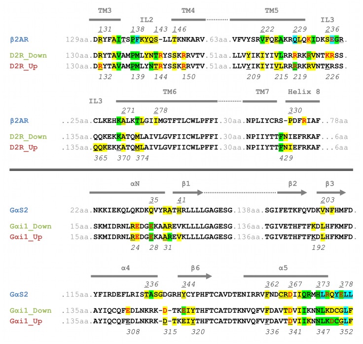
Figure 4. Alignment of the amino-acid contacts between receptors and G-proteins.
Individual alignments for the receptors and the G-proteins are shown. A colored background indicates that the residue forms contacts to other amino acids (yellow: 1 or 2 contacts; green: 3 or 4 contacts; blue: at least 5 contacts). Red letters indicate residues involved in ionic interactions, whereas dotted underlines indicate contacts present in the crystal structure of β2AR-Gαs.

