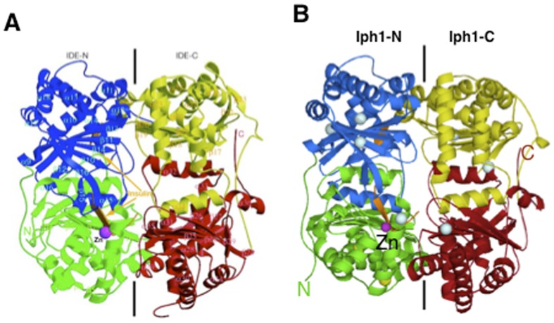
Figure 2. Structures of hIDE and model of Iph1 in complex with the insulin B chain.
A) Crystallographic structure of the human IDE-E111Q–insulin B chain complex (PDB code 2G56) [48]. Domains 1, 2, 3 and 4 are colored green, blue, yellow and red, respectively. The Zn2+ ion and insulin B chain are colored magenta and orange, respectively. B) Model of an insulin B chain/Iph1 complex. The four domains of Iph1 are drawn in the same orientation and color codes as in A. The two Cys conserved in hIDE are in yellow, the remaining Cys residues are in cyan.

