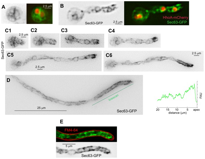
Figure 2. Changes in the ER in swollen conidia, germlings and mature hyphae.
A. Swollen conidium after the first mitotic division. Sec63-labelled ER is shown on the left in inverted grey contrast. The right image is a merge of the Sec63-GFP (green) and HhoA-mCherry (that labels nuclei/chromatin) channels. B. A germling, with fluorescent markers displayed as in A. C. Sec63-GFP ER (inverted contrast) in germlings imaged at different stages after polarity establishment (all images displayed at the same magnification). Peripheral ER strands concentrated near the tip are visible at the stages shown in C3 through C6. The prominence of the tip pm-associated ER strands increases with the length of the germtube. D. Long hypha. A linescan of the Sec63-GFP signal across the indicated line is shown on the right (FAU, fluorescence arbitrary units). E. Cortical ER strands do not overlap with the plasma membrane, stained with FM4-64.

