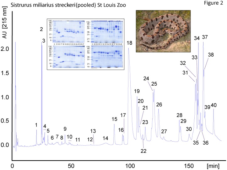
Figure 2. The venom proteome of S. m. streckeri.
The proteins from 2 mg of pooled crude venom were fractionated on a C18 column as described in the Experimental section. HPLC fractions were collected manually and analyzed by SDS-PAGE (insert; under non-reduced (upper panel) and reducing (lower paner) conditions), N-terminal sequencing, and molecular mass determination by ESI-MS or SDS-PAGE. Protein bands excised from SDS-polyacrylamide gel were identified by tryptic peptide mass fingerprinting and CID-MS/MS. The results are listed in Table 1.

