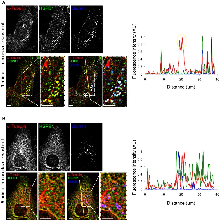
Figure 3. HSPB1 colocalizes to non-centrosomal formation sites only at early stages of repolymerization.
Naive HeLa cells stained for HSPB1, α-tubulin and the Golgi apparatus marker Giantin at (A) 1 min and (B) 5 min after nocodazole washout. Graphs represent line intensity plots for the lines drawn in the corresponding images. Note that HSPB1 does not colocalize with the centrosome (circled in yellow). 3D image deconvolution was applied on the image stacks in (A–B). Scale bar = 5 µm.

