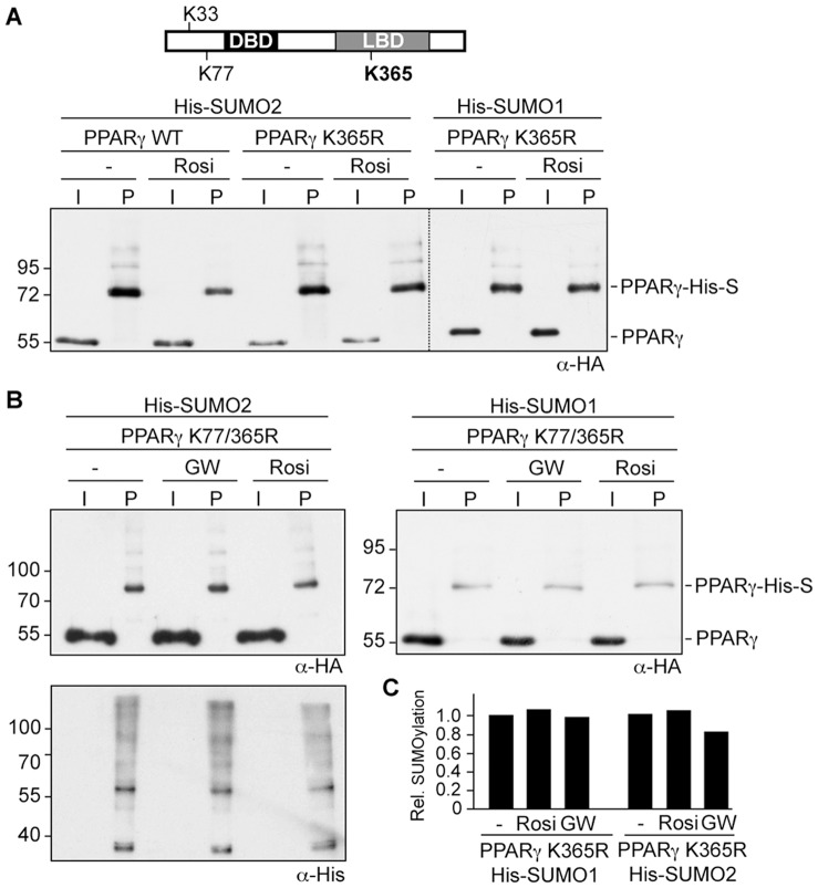
Figure 4. Lysine 365 within the LBD is essential for ligand-induced reduction of PPARγ SUMOylation.
(A) (B) and (C) The PPARγ K365R (A) and PPARγ K77/365R (B) mutants were transfected in HEK293 cells and analyzed for His-SUMO2 and His-SUMO1 modification in the absence and presence of ligands as outlined in the legend to Figure 1. The blot shown in the upper left panel of figure 4B was re-probed with an anti-His antibody to control for loading. (C) Summary of quantitative Western blot analysis. SUMOylation of the PPARγ K365R mutant in the absence or presence of rosiglitazone or GW1929 was analyzed by an independent quantitative Western blot analysis using fluorescence-labeled secondary antibodies. The values obtained for SUMOylated PPARγ K365R relative to the input signal in the absence of ligands were arbitrarily set to 1.

