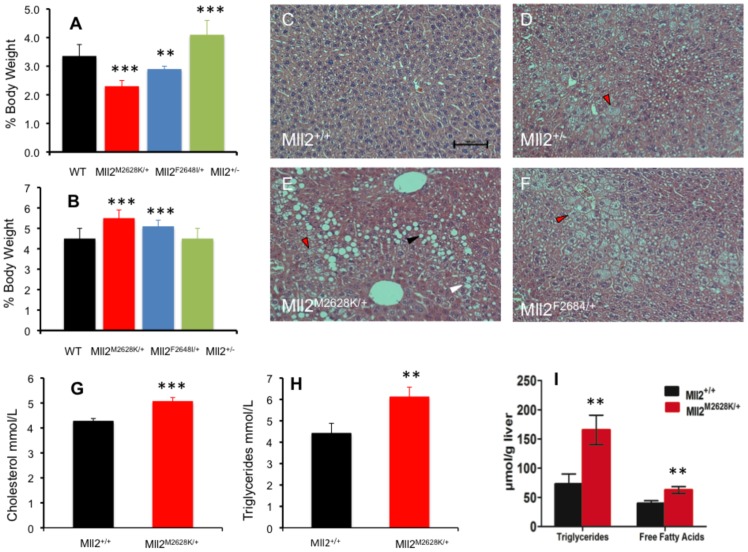
Figure 5. Biochemical and Histological analysis of Mll2 mutants; NAFLD and dyslipidaemia.
Cohorts of mice were culled at 19 weeks of age. A: Epididymal fat pad weights normalized for body weight, Mll2M2628K/+ (N = 10) and Mll2F2648I/+ (N = 7) cohorts exhibited abnormal peripheral fat deposition with reduced fat pads compared to wild-type littermate (N = 23) or Mll2+/− (N = 5). B: Liver weight normalized for body weight, Mll2M2628K/+ (N = 10) and Mll2F2648I/+ (N = 7) cohorts show hepatomegaly. C–D: Histological analysis of H&E stained liver sections demonstrated features consistent with mild NAFLD in all Mll2 mutant and knockout lines (Figure 6C, D & E) with significant increases in macrovesicular steatosis (black arrow), microvesicular steatosis (red arrows) and ballooning hepatocyte degeneration (white arrow), compared to wild-type littermates. Biochemical analysis of plasma showed elevated Cholesterol (G) and Triglycerides (H). I: The steatosis was confirmed biochemically as liver triglycerides and liver free fatty acids were significantly increased in the M2628K mutation. The data represent the mean of 6 animals of each genotype class ±SEM * p<0.05, **p<0.01, ***p<0.001, student's t-test.

