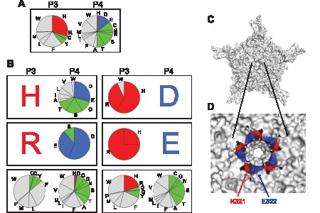
Figure 4. Peculiar positive-negative and neutral-neutral amino acid combinations at P3 and P4 in the 143 viable VP heptapeptides.

(A) Amino acid compositions of all the 143 viable VP heptapeptide mutants. (B) Various amino acid combinations at P3 and P4 positions in the viable VP heptapeptide mutants. Left panels show P3/P4 combinations when histidine (H, upper left), arginine (R, middle left) or a non-charged amino acid (lower left) is at P3. Right panels show P3/P4 combinations when aspartic acid (D, upper right), glutamic acid (E, middle right) or a non-charged amino acid (lower right) is at P4. (C) Topological location of amino acid residues at P3 (K321, red) and P4 (E322, blue) in the wild type AAV2 capsid VP3 is shown on a VP3 pentamer viewed down an icosahedral five-fold symmetry axis at the center. E322 is partially exposed on the outer surface near the five-fold pore while K321 is barely seen from the outside of the capsid. (D) A close-up view of K321 and E322. Ten amino acids (from V323 to I332) forming the outer ridge around the five-fold pore are removed to make K321 and E322 visible. Five pairs of K321 and E322 form a ring in the five-fold channel wall. Panels C and D are created using PyMOL.
