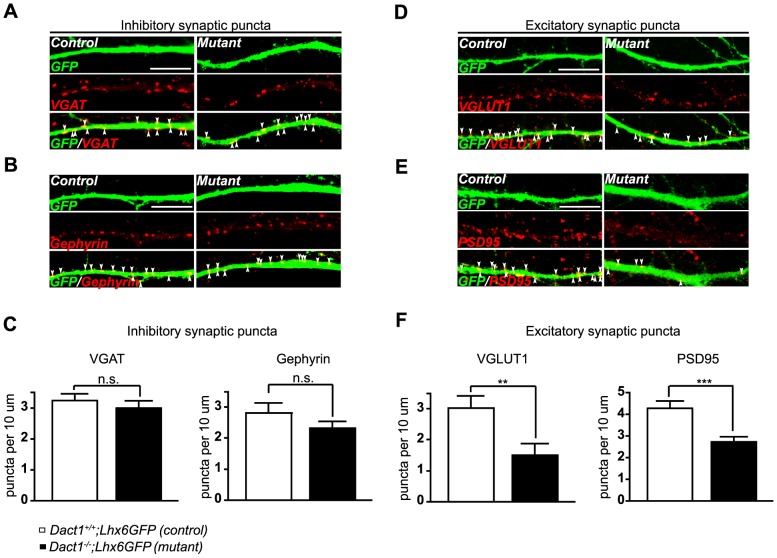
Figure 3. Cortical interneurons from constitutive Dact1 mutant mice have fewer excitatory synapses on primary dendrites.
Primary cortical cultures were prepared from postnatal day 0 Dact1−/−;Lhx6GFP (right) and control (left) brains, fixed at day in vitro 15, and synaptic puncta counted along GFP labeled primary dendrites from the cell soma to the first major branch point (arrowheads). Inhibitory synaptic puncta were visualized using antibodies against VGAT (presynaptic, A) and Gephyrin (postsynaptic, B) with each marker counted irrespective of co-localization with the other; C Quantification per 10 µm of primary dendrite length in control (open bars) and constitutive Dact1 mutant neurons (closed bars). Excitatory synaptic puncta were visualized using antibodies against VGLUT1 (presynaptic, D) and PSD95 (postsynaptic, E) with each marker counted irrespective of co-localization with the other; F Quantification per 10 µm of primary dendrite length. Data shown are mean ± sem of at least 3 independent experiments, collected from at least 3 mice per genotype, 10–15 neurons per animal. **p<0.01; ***p<0.001; n.s., not significant. Scale bars = 10 µm.

