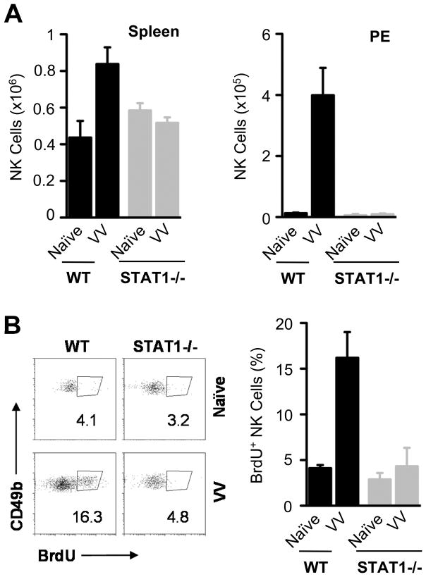
FIGURE 2.
STAT1 −/− mice have decreased NK cell numbers. WT and STAT1 −/− mice were infected with 2 × 10⁶ pfu of VV intraperitoneally or left uninfected (Naïve). Mice were sacrificed 36 hr after infection and NK cells were analyzed in the spleen and in peritoneal cavity exudates (PE). Also, NK cell proliferation in PE was assessed by BrdU incorporation. (A) The mean cell numbers ± SD of CD49b+CD3− NK cells are shown (n = 6). (B) FACS plots showing the percentage of NK cell proliferation by BrdU staining among CD49b+CD3− NK cells (left panel); the mean percentage of NK cell proliferation ± SD among CD49b+CD3− NK cells is indicated (right panel)(n = 4). Data shown is representative of two independent experiments.

