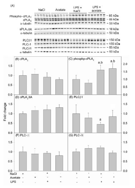
Figure 1. Phospholipases levels in LPS-stimulated BV-2 cell cultures.
Western blot analysis was performed to show changes in the levels of cPLA2 phosphorylation and total cPLA2, sPLA2 IIA, PLCβ1, PLCγ1 and PLCδ1 protein levels in BV-2 microglial cell cultures stimulated with LPS and/or treated with acetate. Panel A shows representative images of the Western blots. Panels B, D, E, F and G show the optical densities total cPLA2, sPLA2 IIA, PLCβ1, PLCγ1 and PLCδ1 normalized to the loading control α-tubulin. Panel C shows the optical density of phosphorylated cPLA2 normalized to total cPLA2. Bars represent means ± SD where statistical significance was set at p ≤ 0.05. Abbreviations are: a = compared to NaCl-treated group and b = compared to sodium acetate-treated group (n = 6 per group).

