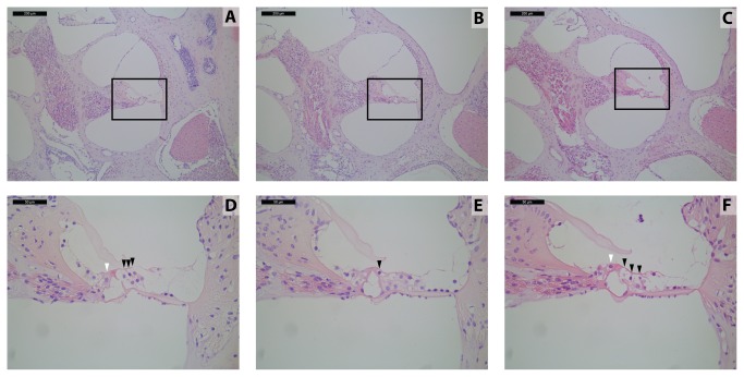
Figure 4. Deaf11 and Deaf13 cochlear hair cells are morphologically abnormal.
Middle cochlear turn sections at 8 weeks of age; Upper panels 100x magnification, scale bar 200 µm; Lower panels 400x magnification, scale bar 50 µm; A, D) Atp2b2 +/+ B, E) Atp2b2 Deaf11/Deaf11 C, F) Atp2b2 Deaf13/Deaf13; White arrowheads point to inner hair cells; Black arrowheads point to outer hair cells; Each section is representative of 4 cochleae.

