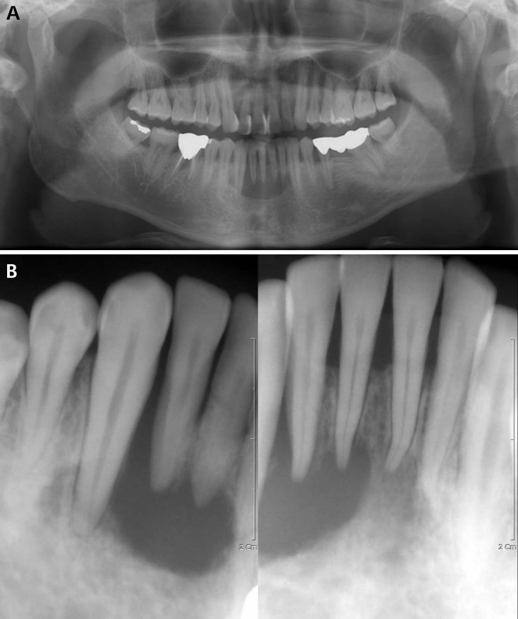
Fig. 1.
A. A panoramic radiograph shows a radiolucent lesion with a well-defined margin from the left lower lateral incisor to the right lower canine. B. A periapical radiograph reveals a radiolucent lesion with a non-corticated border and beveled edges. The distal part of the right lower lateral incisor shows a "floating tooth" appearance.

