Fig. 5.

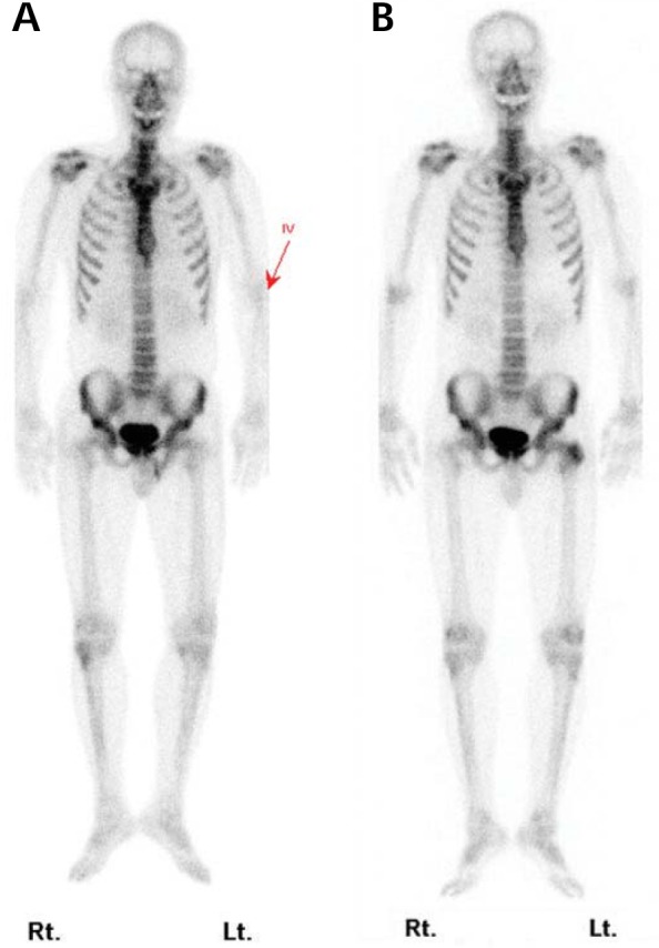
Bone scintigraphy. A. The initial study shows normal uptake of the radiotracer in the trochanteric portion of the left femur. B. The one year follow-up study demonstrates an uptake lesion in the left femur.

Bone scintigraphy. A. The initial study shows normal uptake of the radiotracer in the trochanteric portion of the left femur. B. The one year follow-up study demonstrates an uptake lesion in the left femur.