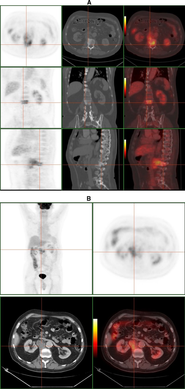
Fig. 10.
F-18 FDG PET–CT study of a 50-year-old man with tuberculous spondylitis. a High FDG uptake located in L2 combined with bone sclerosis on CT scan. b There is a extraosseous fixation of the tracer, that corresponds to a paraspinal abscess according to CT image (courtesy of Dr. Lorenzo-Bosquet)

