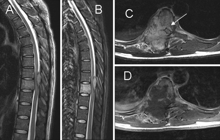
Fig. 4.
Paradiskal pattern. a, b Sagittal T2-weighted imaging and STIR show hyperintense signal in the T9 vertebral body, suggesting bone marrow edema. c, d Axial T2- and T1-postcontrast weighted imaging show paradiskal involvement and infection spreading to the epidural space (arrow) with spinal cord compression

