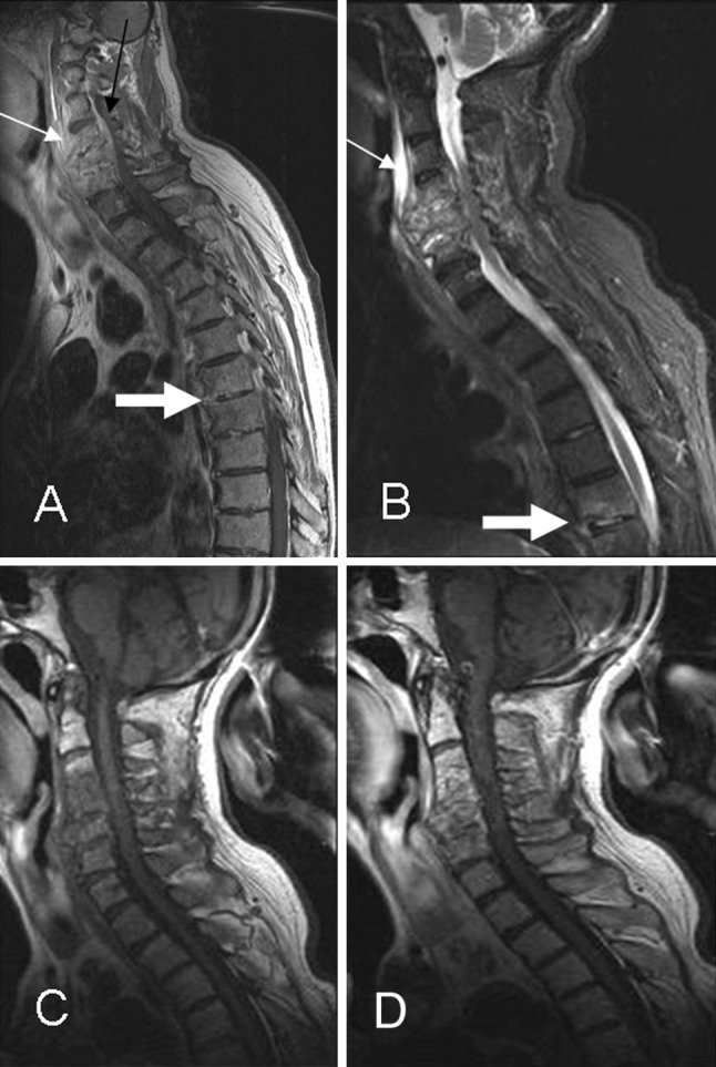
Fig. 9.

Multisegmentary infection. a, b Sagittal T1-postcontrast weighted and STIR imaging show signs of advanced cervical infection with a large prevertebral abscess (thin arrow) and early skip lesion with epidural enhancement involving T6 and T7 levels (thick arrow). c, d Follow-up MRI 2 months later shows persistence of contrast enhancement in the cervical vertebral bodies with resolution of the prevertebral abscess
